Objective: Optical detection systems have potential to address challenges in cervical cancer care. Compact, inexpensive light sources, sensors, and wireless internet connectivity are ubiquitous. It is possible to build mobile multi-spectral imaging systems that acquire data at the point of care, and perform computations in the cloud to estimate blood content, oxygen saturation, and light scattering in the cervix. Over time, this additional information would enable better triage at the point of care.
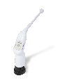
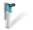
Methods: A novel mobile multi-spectral imaging system was built around a tablet form factor, with a 13-LED illumination board and an area-scan camera. Image frames corresponding to each wavelength were recorded. Each multi-spectral cervical data set was captured in under five seconds and uploaded to the cloud for computational analyses. Pixel wise calculations of blood content, oxygen saturation, and scattering were made in the cloud and returned to the clinician. Sites of cervical biopsies were tracked in software; spectral output were compared to histopathology. The protocol was for this pilot study was approved by the institutional review board. Results: Images of blood content, oxygen saturation, and scattering were obtained. The estimated values were within the range expected for epithelial tissues. Abnormal measurements of blood content and scattering spatially correlated with biopsy sites. Analyses were done in near real time.
Conclusions: Full resolution images of blood content and oxygen saturation of cervical tissues hold great potential to supplement current cervical cancer screening methods. Future upgrades will reduce the device size and analysis time.